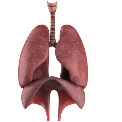
The respiratory system has many different parts that work together to help you breathe. Your mouth and nose pull air from outside your body into your respiratory system. There are sinuses, which are hollow areas between the bones in your head that help regulate the temperature and humidity of the air that is inhaled. The pharynx is a tube that delivers air from your mouth and nose to the trachea, a passage connecting your throat and lungs. Bronchial tubes are at the bottom of the trachea that connect into each lung. The lungs remove oxygen from the air and pass it into your blood. The bloodstream delivers oxygen to all your organs and other tissues
But that's not all! Muscles and bones help move the air you inhale into and out of your lungs. Some of the bones and muscles in the respiratory system include the diaphragm, which helps your lungs pull in air and push it out. The ribs surround and protect the lungs and heart. 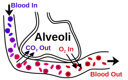
Other important components of the system are:
Using some common materials, we can build a lung model to help us learn how they function. Our model can also help us remember some important structures that are a necessary part of our respiratory system.
CHECK OUT MY TIKTOK VIDEO DEMONSTRATION BELOW, AND THEN READ ON FOR MORE DETAILS!
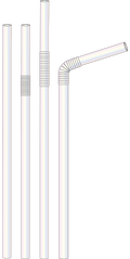
Materials
Procedure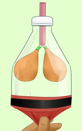
Model Breathing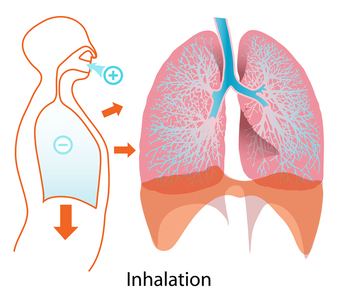
Gently pull down on the diaphragm. This should cause air to flow into the lungs. Allow the diaphragm to return/move upward and the air from the lungs will be expelled. Scientific Explanation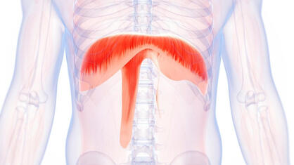
The diaphragm is a strong muscle that expands and contracts. As it does so, it causes your lungs to fill with air and then empty out again.
The movement of the balloon-diaphragm matches your own breathing – when you breathe in, your lungs fill with air just like the balloons inside the bottle do. That’s because the diaphragm expanded making room for air inside the lungs. When you breathe out, your diaphragm contracts (or squeezes in) pushing all the air out of your lungs. In your model, as the diaphragm balloon is pulled it creates more space inside the bottle. Air then comes down the trachea straw and fills the lung balloons with some air to fill the space! When you let go of the diaphragm balloon, the space no longer exists, so the air from the lung balloons is expelled making them deflate. Imperfect lung model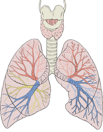
Although this lung model is fabulous, there are some characteristics it does not share with real lungs. Human lungs aren’t the same size. To accommodate the heart, the right lung is larger than the left lung. Also, lungs have lobes. The right lung has three and there is two lobes in the left. Thin sacs called pleura surround each lobe and separate them from the chest wall. Think About It
After student models have been completed, discuss respiratory function with students. Have them place their hands on their chest breath in and out. Ask them to explain what happens to their chest when they exhale and inhale and why it does so. Other questions you could ask include:
0 Comments
|
AuthorGertrude Katz has spent over 30 years teaching K-12 public school students all major subjects. She has taught biology and education at the college level. The majority of her career has been spent instructing biology at the secondary level. Categories
All
|

 RSS Feed