
Whether you're trying to impart some information about Human Body Systems, you're discussing homeostasis, it's late October and you need a holiday-specific lesson, or you're looking for some random factoids to fill time between units, this post is chock-full of classroom gold! So, don't be a bone-head! Read on for the top ten teachable bone facts!
#1: Bones have purpose! They provide the structure for our bodies. They are made of connective tissue reinforced with calcium and specialized cells. Most bones also contain bone marrow, where blood cells are made.
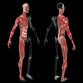
Bones work with muscles and joints to hold our body together and support freedom of movement. This is called the musculoskeletal system. The skeleton supports and shapes the body and protects delicate internal organs such as the brain, heart, and lungs.
Bones contain most of our body’s calcium supply. The body is constantly building up and breaking down bone tissue as required.
The Skeleton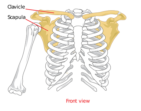
#2: The human skeleton is made up of 206 bones. The most common ones are listed below. Bone up on the scientific names to show everyone you're not a numb-skull!
Bone Types
#3: There are four different types of bone in the human body:
Bone Tissue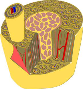
#4: The different layers of bone tissue include:
Bone Cells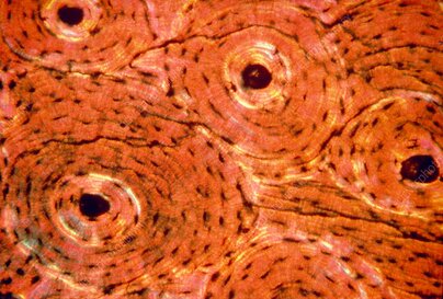
#5: Our body is constantly remodeling its skeleton by building up and breaking down bone tissue as required. As a result, each bone is rebuilt from scratch about every decade. The bone cells involved in this process include:
Bone Density
#6: Many factors work together to ensure the strength and health of bones. Bone density relies on:
Joints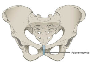
#7: Joints hold the skeleton together and support movement. Joints may be categorized by function (range of motion) or by structure (material). Some joints are immovable, such as skull sutures and the articulations between the teeth and jaws. Other joints allow slight movement like the membrane between the tibia and the fibula and the pubic symphysis of the pelvic girdle. Joints allowing full movement include bone articulations in the limbs like the elbow, shoulder, and ankle.
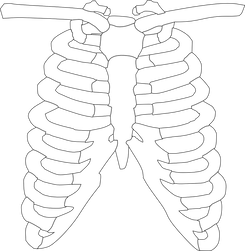
#8: Joints may be structurally either fibrous, cartilaginous, or synovial. Fibrous joints may be immovable (like the sutures that connect skull bones and gomphoses that connect teeth to the jaws) or slightly movable (like the syndesmosis between the tibia and fibula called the interosseous membrane). Examples of cartilaginous joints are those between the first pair of ribs and the sternum, as well as the pubic symphysis and the vertebrae. 
#9: Synovial joints are characterized by the presence of an articular capsule between the two joined bones. Bone surfaces at synovial joints are protected by a coating of articular cartilage. Synovial joints are often supported and reinforced by surrounding ligaments, which limit movement to prevent injury. 
#10: There are six types of synovial joints:
Bonus teachable activity!!
Make a skeleton out of paper towel and toilet paper rolls! Check out this easy video to see how you can use this hands-on activity to help students learn about this necessary body system
Watch G. Katz Chronicle TikTok & YouTube Videos for demonstration of how to make your own TP / PT Skeleton Model!
Find Disarticulated Skeleton Bone Cutouts Here!
0 Comments
Leave a Reply. |
AuthorGertrude Katz has spent over 30 years teaching K-12 public school students all major subjects. She has taught biology and education at the college level. The majority of her career has been spent instructing biology at the secondary level. Categories
All
|
 RSS Feed